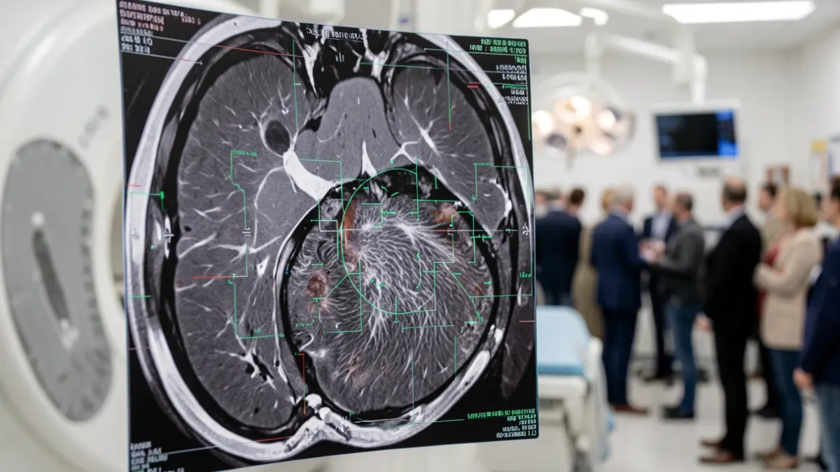
The findings, published in the scientific journal International Journal of Obesity, indicate that the characteristics of belly fat, not just its quantity and location, are associated with heart artery disease. The technique acts as a virtual biopsy, where artificial intelligence algorithms extract hundreds of tissue characteristics, such as density and texture, invisible to the naked eye.
The team analyzed scans from 1,736 adults without known coronary artery disease, focusing on subcutaneous fat, visceral fat, and total abdominal fat. Scientists identified a “pro-atherogenic signature,” a fat with a more heterogeneous texture, associated with the presence of calcification in the coronary arteries, a marker of atherosclerosis and increased heart attack risk.
“When abdominal fat becomes very heterogeneous in its texture, it acts as a warning sign for the presence of calcification in the coronaries,” states the FMUP researcher.
The study also revealed pronounced differences in abdominal fat structure between men and women. “Fat is not all the same. What makes it dangerous is not so much where it is, but the characteristics it presents,” explains Jennifer Mancio.
Radiomics could be applied on a large scale to understand fat quality, making the process fully automated and eliminating the need for manual segmentation. According to the researcher, this analysis involves no additional costs or radiation for patients, as it utilizes CT scans already requested for other reasons. In the future, it could help identify earlier who has fat characteristics with a higher risk of coronary calcification and personalize the prevention and treatment of individuals with obesity.
The study was conducted as part of the SAFE-CT project and funded by the CUF D. Manuel de Mello Scholarship, from the Fundação Amélia de Mello.